Clec4g Antibody
-
货号:CSB-PA803912ZA01MO
-
规格:¥440
-
促销:
-
图片:
-
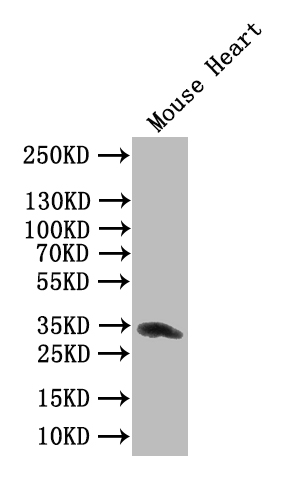
Western Blot
Positive WB detected in: Mouse Heart tissue lysate
All lanes: Clec4g antibody at 1:1000
Secondary
Goat polyclonal to rabbit IgG at 1/50000 dilution
Predicted band size: 34 kDa
Observed band size: 34 kDa -
Western Blot
Positive WB detected in: Mouse Heart tissue lysate
All lanes: Clec4g antibody at 1:1000
Secondary
Goat polyclonal to rabbit IgG at 1/50000 dilution
Predicted band size: 34 kDa
Observed band size: 34 kDa -
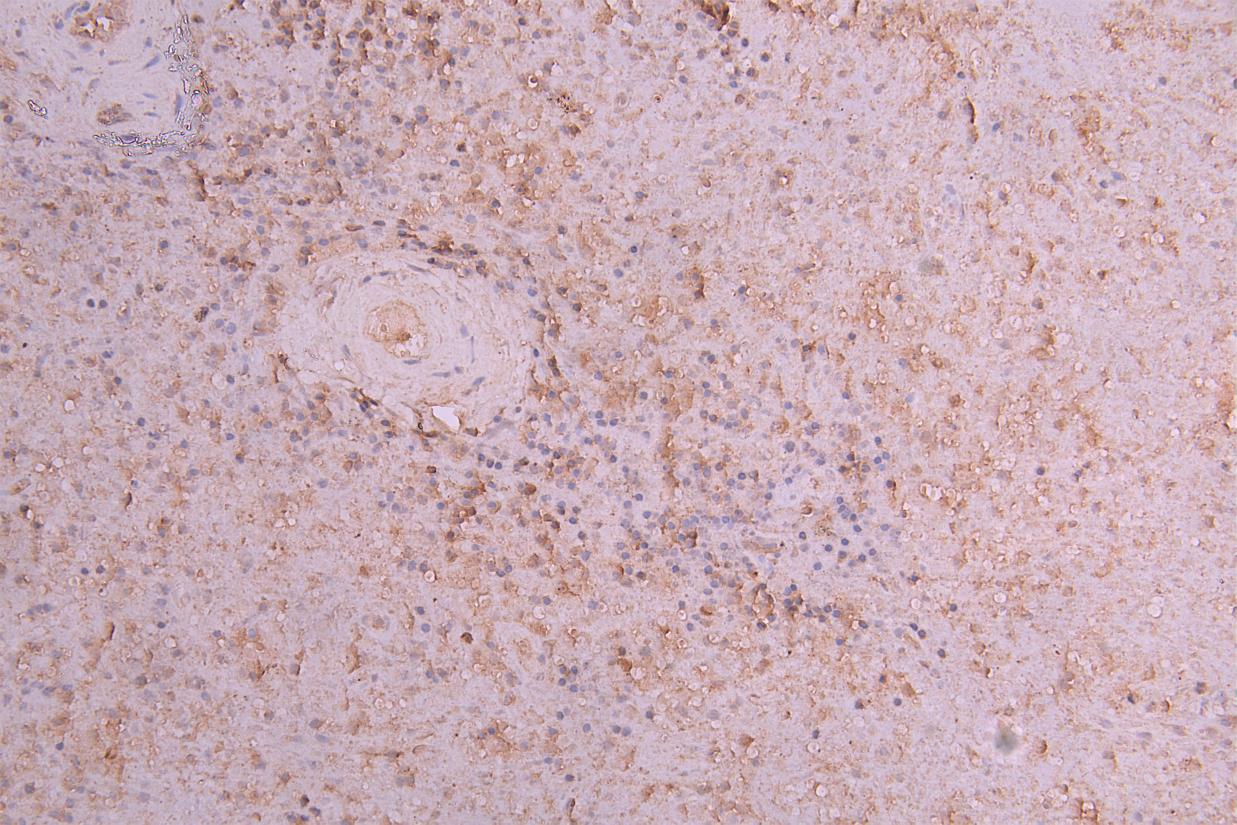
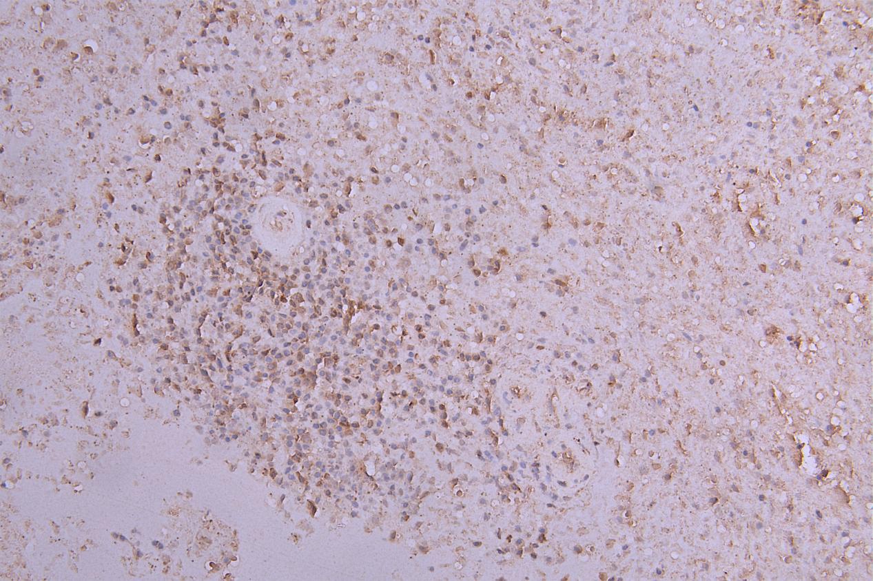
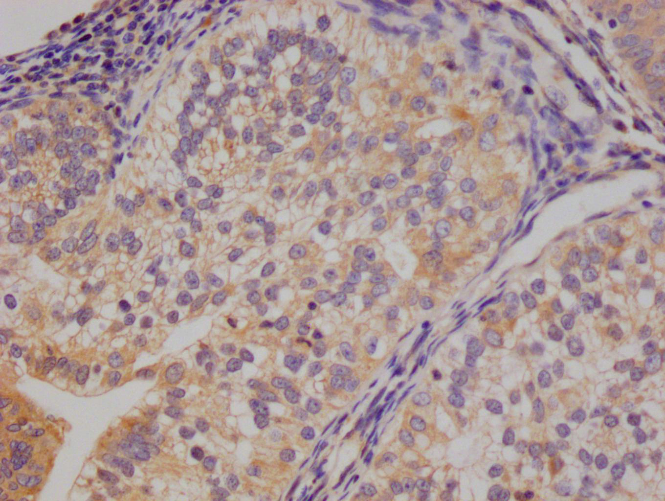
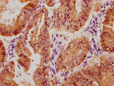
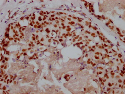
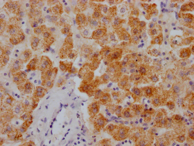
IHC image of CSB-PA803912ZA01MO diluted at 1:66 and staining in paraffin-embedded human spleen tissue performed on a Leica BondTM system. After dewaxing and hydration, antigen retrieval was mediated by high pressure in a citrate buffer (pH 6.0). Section was blocked with 10% normal goat serum 30min at RT. Then primary antibody (1% BSA) was incubated at 4°C overnight. The primary is detected by a Goat anti-rabbit polymer IgG labeled by HRP and visualized using 0.05% DAB.
-
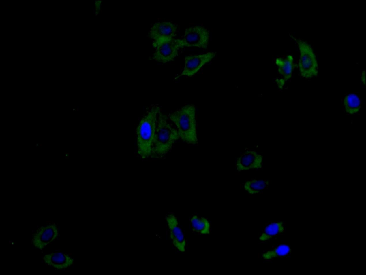
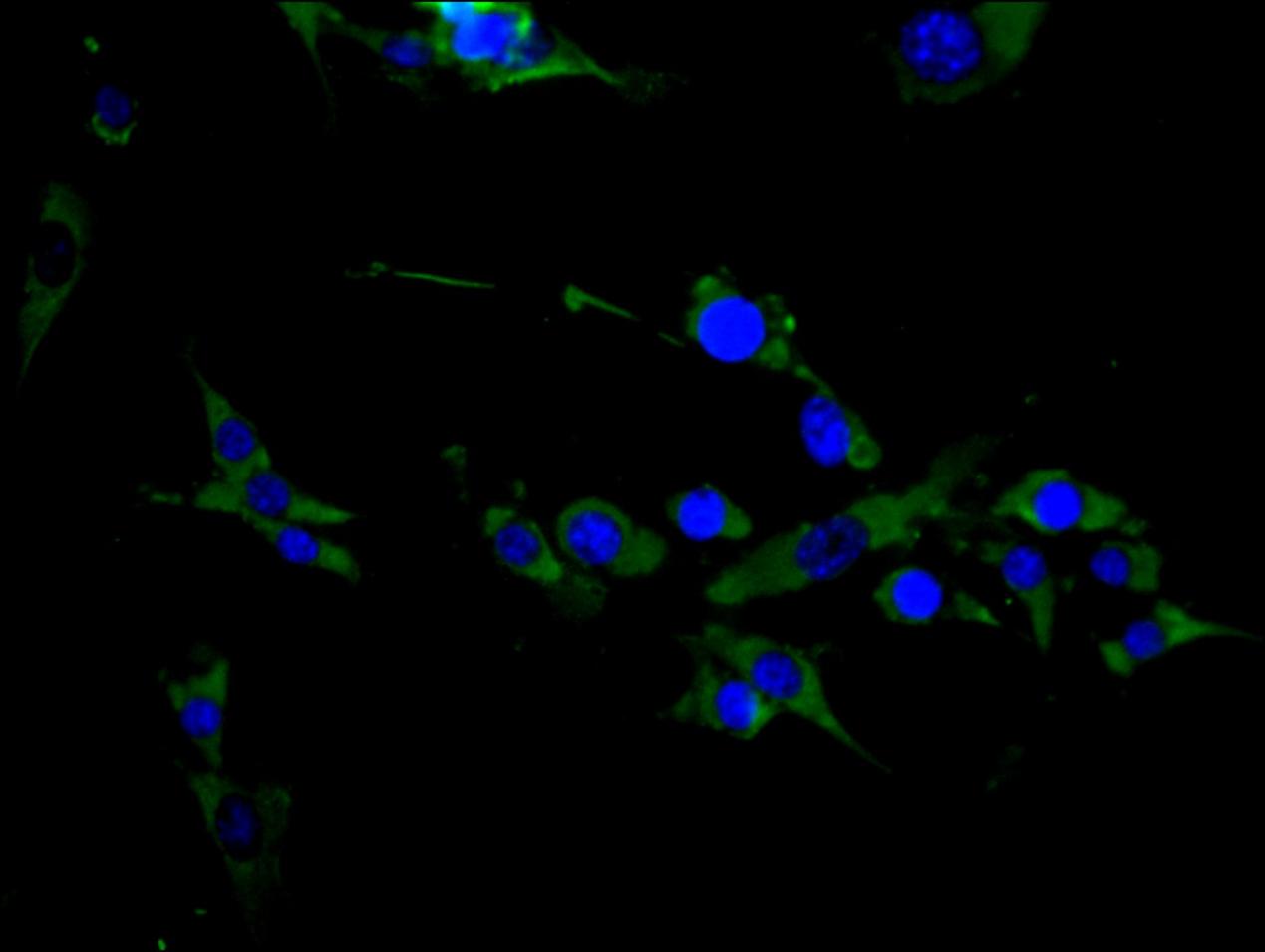
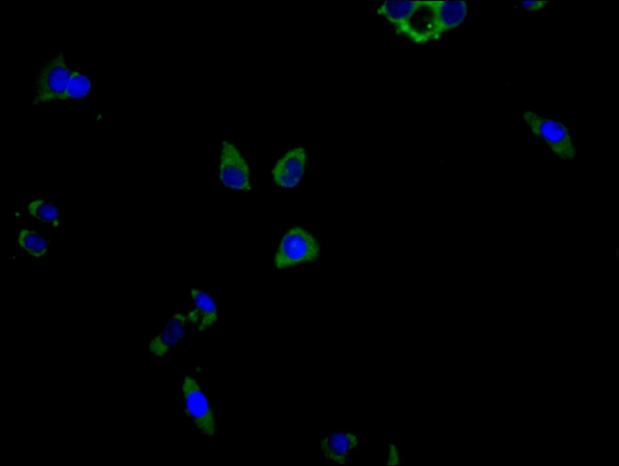
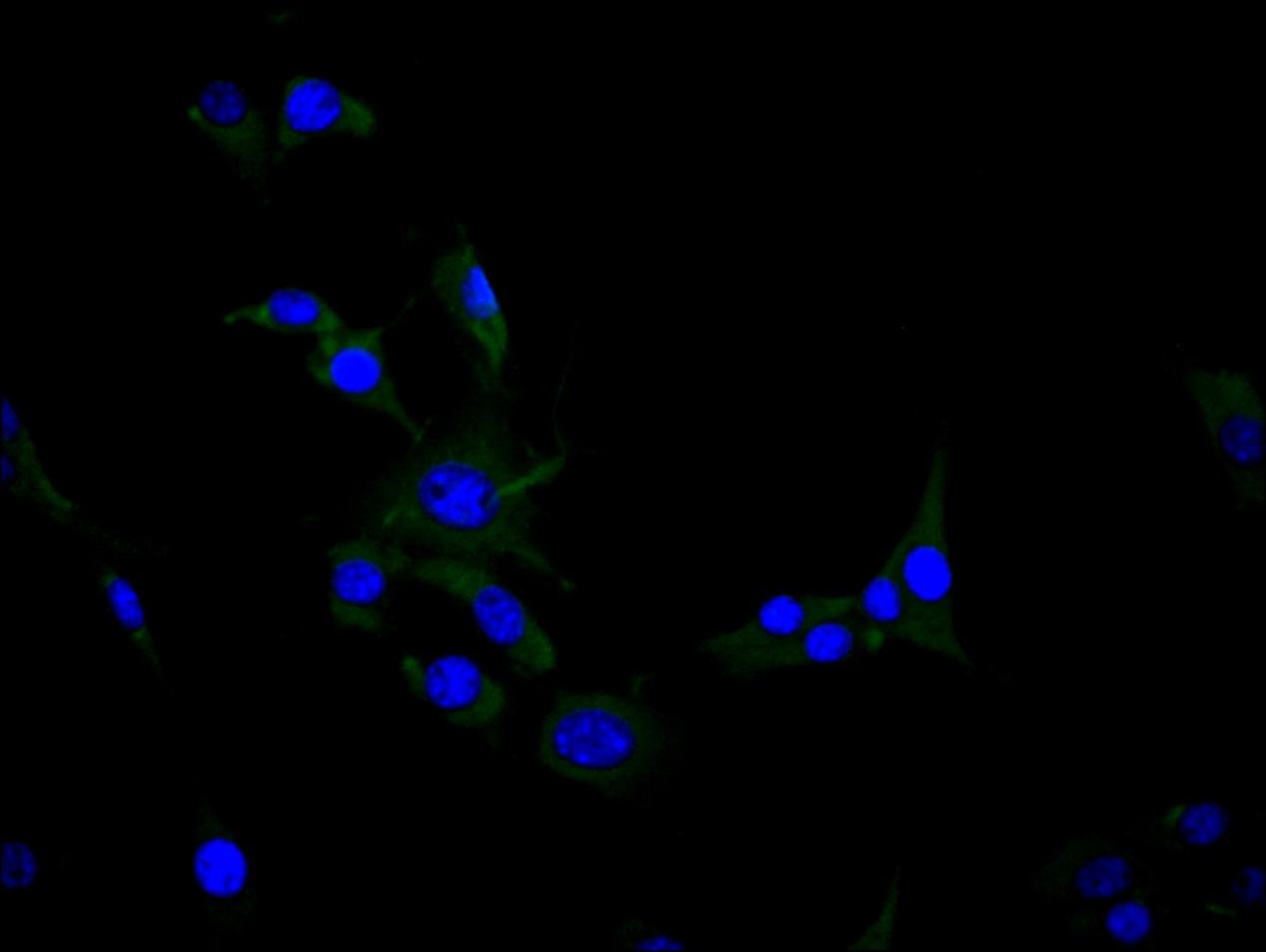
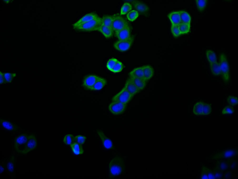
Immunofluorescence staining of Hela cell with CSB-PA803912ZA01MO at 1:30, counter-stained with DAPI. The cells were fixed in 4% formaldehyde and blocked in 10% normal Goat Serum. The cells were then incubated with the antibody overnight at 4C. The secondary antibody was Alexa Fluor 488-congugated AffiniPure Goat Anti-Rabbit IgG(H+L).
-
Immunofluorescence staining of NIH/3T3 cell with CSB-PA803912ZA01MO at 1:30, counter-stained with DAPI. The cells were fixed in 4% formaldehyde and blocked in 10% normal Goat Serum. The cells were then incubated with the antibody overnight at 4C. The secondary antibody was Alexa Fluor 488-congugated AffiniPure Goat Anti-Rabbit IgG(H+L).
-
-
其他:
产品详情
-
产品名称:Rabbit anti-Mus musculus (Mouse) Clec4g Polyclonal antibody
-
Uniprot No.:Q8BNX1
-
基因名:Clec4g
-
别名:C-type lectin domain family 4 member G, Clec4g
-
宿主:Rabbit
-
反应种属:Mus musculus
-
免疫原:Recombinant Mus musculus Clec4g protein (52-294aa)
-
免疫原种属:Mus musculus (Mouse)
-
标记方式:Non-conjugated
-
克隆类型:Polyclonal
-
抗体亚型:IgG
-
纯化方式:Antigen affinity purification
-
浓度:It differs from different batches. Please contact us to confirm it.
-
保存缓冲液:Preservative: 0.03% Proclin 300
Constituents: 50% Glycerol, 0.01M PBS, pH 7.4 -
产品提供形式:Liquid
-
应用范围:ELISA, WB, IHC, IF
-
推荐稀释比:
Application Recommended Dilution WB 1:500-1:5000 IHC 1:50-1:200 IF 1:30-1:100 -
Protocols:
-
储存条件:Upon receipt, store at -20°C or -80°C. Avoid repeated freeze.
-
货期:Basically, we can dispatch the products out in 1-3 working days after receiving your orders. Delivery time maybe differs from different purchasing way or location, please kindly consult your local distributors for specific delivery time.
相关产品
靶点详情
-
功能:Binds mannose, N-acetylglucosamine (GlcNAc) and fucose, but not galactose, in a Ca(2+)-dependent manner.
-
基因功能参考文献:
- This study shows that C-type lectin LSECtin, a member of the DC-SIGN family, is a novel liver regulator for natural killer cells (NK cells). LSECtin could bind to NK cells in a carbohydrate-dependent manner and could regulate the number of hepatic NK cells. PMID: 27184407
- Data show that C-type lectin-like domain family 4, member g (Clec4g) suppresses beta-Site amyloid precursor protein cleaving enzyme-1 (BACE1)-mediated amyloid-beta (Abeta) peptides generation. PMID: 25957769
- When expressed in B16 melanoma cells, LSECtin promoted tumor growth, whereas its blockade slowed tumor growth in either wild-type or LSECtin-deficient mice. PMID: 24769443
- LSECtin-mediated signaling up-regulates the threshold of CD4(+) T cell activation via tuning the expression of Cbl-b. PMID: 22884358
- LSECtin may facilitate the reduction of liver inflammation at the cost of delaying virus clearance, an effect that might be hijacked by the virus as an escape mechanism. PMID: 23487419
- The studies provide a basis for using mouse LSECtin, and knockout mice lacking this receptor, to model the biological properties of the human receptor. PMID: 21257728
- Liver sinusoidal endothelial cell lectin, LSECtin, negatively regulates hepatic T-cell immune response. PMID: 19632227
显示更多
收起更多
-
亚细胞定位:Cell membrane; Single-pass type II membrane protein.
-
数据库链接:
KEGG: mmu:75863
STRING: 10090.ENSMUSP00000059574
UniGene: Mm.109183
Most popular with customers
-
-
YWHAB Recombinant Monoclonal Antibody
Applications: ELISA, WB, IF, FC
Species Reactivity: Human, Mouse, Rat
-
Phospho-YAP1 (S127) Recombinant Monoclonal Antibody
Applications: ELISA, WB, IHC
Species Reactivity: Human
-
-
-
-
-


